Modeling physiology using dynamic medical imaging and machine learning
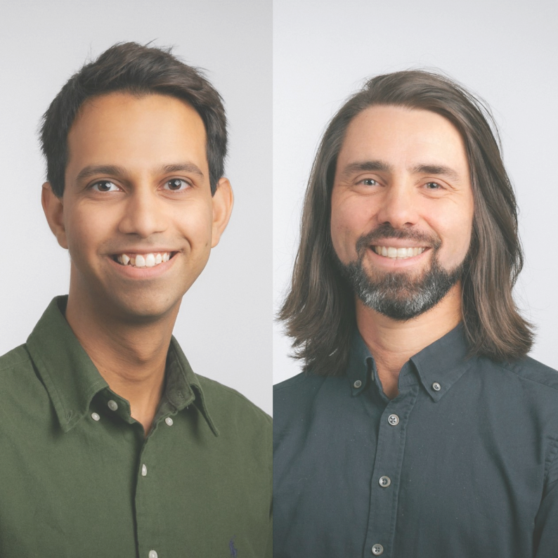
Kartikay Tehlan and Prof. Dr. Thomas Wendler
Universitätsklinikum Augsburg
Details

Dynamic medical images typically capture how contrast media distributes inside the body over seconds to minutes. In this talk, we show how these three dimensional movies can be viewed as time varying biosignals at each location in the body, enabling us to measure physiology rather than just anatomy. We build simple "compartment" models - think of them as flow charts for where the contrast media can go - that summarize how the injected agents move between tissues and cells. With these models, we apply mathematics to link what the scanner sees to what tissues are doing. Because the equations are large and noisy, we use modern AI methods to solve them efficiently and stably, producing interpretable parametric images that map biological processes across the body. We will illustrate this approach using dynamic PET (positron emission tomography) with a tiny amount of radioactive sugar (FDG) as contrast media. By estimating parameters that describe how fast FDG is taken up and retained by tissues, the resulting parametric images can help distinguish inflammation from malignant lesions and provide a sensitive tool for detecting very early tumor regression during therapy. The goal is a plain language walkthrough: how dynamic imaging, simple physiological models, and AI come together to deliver clearer, more actionable pictures of health and disease. |
